Muscles Of The Chest Abdomen And Thigh (Superficial Dissection) : Contents Preface Acknowledgments List Of Clinical Blue Boxes List Of Tables Figure Credits 1 Overview And Basic Concepts Approaches To Studying Anatomy Regional Anatomy Systemic Anatomy Clinical Anatomy Anatomicomedical Terminology : The thorax is located in the upper trunk, defined anteriorly by the sternum bone, laterally by the ribs, and later by the spine.
Muscles Of The Chest Abdomen And Thigh (Superficial Dissection) : Contents Preface Acknowledgments List Of Clinical Blue Boxes List Of Tables Figure Credits 1 Overview And Basic Concepts Approaches To Studying Anatomy Regional Anatomy Systemic Anatomy Clinical Anatomy Anatomicomedical Terminology : The thorax is located in the upper trunk, defined anteriorly by the sternum bone, laterally by the ribs, and later by the spine.. Inserts at iliac crest and linea alba 3. Fabian identifying the muscles and landmarks of the abdomen and chest. Want to learn more about it? Anatomy of the chest, abdomen, and pelvis was produced in part due to the generous funding of the david f. Contraction of the diaphragm causes it to descend towards the abdomen, increasing the space of the thoracic cavity and expanding the lungs.
Pain that increases with movement of the chest or upper spine. Conclusion moving down the trunk of the cat from the chest to the abdomen, i was able to identify the latissimus dorsi, internal oblique, transverse abdominus, rectus. The thigh is the area between the hip and the knee joint. It works to move forelimb towards the chest. Inferior border of each rib.
Abdominal Muscles Location And Function from www.verywellfit.com The pain tends to persist and it worsens with activity. The space between pelvis and chest is called abdomen which is commonly known as belly. The single bone in the thigh region is called the femur. Sound all actions and name the symptoms you are testing. Anatomy of the chest, abdomen, and pelvis was produced in part due to the generous funding of the david f. It consists of areolar tissue containing in its meshes much fat, and may be separated into two or more layers, between which are found the superficial vessels and nerves. For some smaller muscle observations, larger. The nervous system consists of the brain and spinal cord, nerves, ganglia and receptors.
Related online courses on physioplus.
It consists of areolar tissue containing in its meshes much fat, and may be separated into two or more layers, between which are found the superficial vessels and nerves. Conduct a general examination and examination of the abdomen and determine all pathological symptoms. Reset help axial muscles appendicular muscle reclus sheath tensor fascia latae external oblique platysma reclus femoris serralus anterior. Find out the patient's complaints, collect a history of the disease. Pain that increases with movement of the chest or upper spine. Inferior border of each rib. Ɪkˈstrɛmɪti] нижняя конечность upper extremity ˈʌpə(r) ɪkˈstrɛmɪti верхняя конечность thigh θai n бедро. Conclusion moving down the trunk of the cat from the chest to the abdomen, i was able to identify the latissimus dorsi, internal oblique, transverse abdominus, rectus. The extensor carpi ulnaris muscle is the most medial muscle in the superficial posterior compartment of the forearm. The pain tends to persist and it worsens with activity. Inserts at iliac crest and linea alba 3. The adductor minimus muscle is a small, variably present muscle in the medial compartment of the thigh. Muscles of the chest, abdomen and thigh (superficial dissection) drag the labels to the appropriate location in the figure.
Contraction of the diaphragm causes it to descend towards the abdomen, increasing the space of the thoracic cavity and expanding the lungs. The abdominal internal oblique muscle. The muscular system consists of the skeletal muscles and their associated structures. Location of the latissimus dorsi muscle: You may feel chest pain anywhere from your neck to your upper abdomen.
Anatomical Position And Directional Terms Anatomy And Physiology from www.registerednursern.com Find out the patient's complaints, collect a history of the disease. In addition to moving the arm and pectoral girdle, muscles of the chest and upper back work together as a group to support the vital process of breathing. Originates from the lower ribs b. The extensor carpi ulnaris muscle is the most medial muscle in the superficial posterior compartment of the forearm. Transverse abdominal compresses abdomen a. Location of the latissimus dorsi muscle: The nervous system consists of the brain and spinal cord, nerves, ganglia and receptors. In the male this layer is continued over the penis.
Muscles of the chest and abdomen— presentation transcript 5 2.
In addition to moving the arm and pectoral girdle, muscles of the chest and upper back work together as a group to support the vital process of breathing. The space between pelvis and chest is called abdomen which is commonly known as belly. Swensen fund for innovation in teaching. The muscle striations, are they easily visible on the cat as they are in the dissection book or are they procedure: In the male this layer is continued over the penis. It is a long, thin, superficial muscle that extends down the length of the thigh in the anterior compartment muscles. Contraction of the diaphragm causes it to descend towards the abdomen, increasing the space of the thoracic cavity and expanding the lungs. While examining the chest, note the shape of the chest, its symmetry (static inspection), type of respiration, participation of the chest wall in hypersthenic chest: The thorax is located in the upper trunk, defined anteriorly by the sternum bone, laterally by the ribs, and later by the spine. Superficial fascia.—the superficial fascia forms a continuous layer over the whole of the thigh; Muscle performance in neck pain assessment and rehab of the deep and superficial neck muscles in the presence of pain powered by physiopedia. The thigh is the area between the hip and the knee joint. When the diaphragm contracts, it pulls the lower wall of the chest cavity down, increasing the volume of the lungs which then fill with air.
Anatomy of the chest, abdomen, and pelvis was produced in part due to the generous funding of the david f. Pain that increases with movement of the chest or upper spine. The adductor minimus muscle is a small, variably present muscle in the medial compartment of the thigh. The oblique muscles run horizontally around the sides of the trunk. Sound all actions and name the symptoms you are testing.
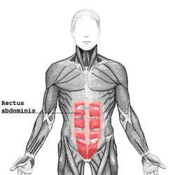
Rectus Abdominis Muscle Wikipedia from upload.wikimedia.org Contraction of the diaphragm causes it to descend towards the abdomen, increasing the space of the thoracic cavity and expanding the lungs. The muscles of the abdomen anterolateral muscles | posterior musclesthe muscles of the abdomen may be divided into anterolateral and below, it passes over the inguinal ligament, and is continuous with the superficial fascia of the thigh. It works to move forelimb towards the chest. Intercostal muscle strains are the most common cause of musculoskeletal chest pain, which people often refer to as a pulled muscle. For some smaller muscle observations, larger. Terminology due to confusion about this muscle over the years it has ended up with a variety of names, which include pars lateralis, adduc. The thorax is located in the upper trunk, defined anteriorly by the sternum bone, laterally by the ribs, and later by the spine. Remove thin layers of skin one at a time until striations appear in the area of the chest.
In the male this layer is continued over the penis.
Muscles of the chest, also called the thorax, include both smooth muscles and skeletal muscles. Conduct a general examination and examination of the abdomen and determine all pathological symptoms. Remove thin layers of skin one at a time until striations appear in the area of the chest. Find out the patient's complaints, collect a history of the disease. The oblique muscles run horizontally around the sides of the trunk. Pain that increases with movement of the chest or upper spine. The superficial back muscles are situated underneath the skin and superficial fascia. Want to learn more about it? The adductor minimus muscle is a small, variably present muscle in the medial compartment of the thigh. For some smaller muscle observations, larger. Terminology due to confusion about this muscle over the years it has ended up with a variety of names, which include pars lateralis, adduc. Transverse abdominal compresses abdomen a. Fabian identifying the muscles and landmarks of the abdomen and chest.
It works to move forelimb towards the chest muscles of the chest abdomen. The superficial back muscles are situated underneath the skin and superficial fascia.

:max_bytes(150000):strip_icc()/external-oblique-muscle-107702789-5bfd926046e0fb002642bdcb.jpg)


0 Komentar